Dinosaurs and Cavemen is a Success!

Greetings, I wanted to take this space to report on a fairly successful event were were able to pull of at Rock Bridge High School. As described previously, members of the Department of Pathology and Anatomical Sciences and the Department of Anthropology (the umbrella term we like is Integrative Anatomy) got together to host a K-12 and beyond outreach event alongside Columbia Public Schools Planetarium. Before I get started I wanted to thank everyone that put so much time and effort into the production and then execution of the event:

Libby Cowgill, Kevin Middleton, Scott Maddux, Dave Dufeau, Carol Ward, Alex Woods, Elizabeth Moffett, Sarah Swartz, Ian George, Henry Tsai, Amy Reynolds Warren, Chet Savage, Rachel Munds, Kaleb Sellers, Elizabeth Lo Presti, Ally McEntire, Ashley Hammond, Rob’yn Johnston, Zack Winkler, and finally the Director of the Planetarium Melanie Knocke.

We had 400 people come see 5, 40 minute long planetarium shows (Earth’s Wild Ride) and the Planetarium collected a little less than $500 in donations. Before and after each show, guests were given the opportunity to take part in a number of activities we developed. The movie touched on numerous parts of Earth’s tumultuous history including the Late Cretaceous Extinction event and the Late Pleistocene glaciation of Europe, hence “Dinosaurs & Cavemen”. The Missourian newspaper had a good write-up with photos. 

We front-loaded the effort on this to develop a number of activities that we can easily employ during future outreach events. As several of the faculty are always trying to figure out how to incorporate their research into broader impacts, it is events like this that not only help us get our chops up, but help expose the community to what is actually going on at Mizzou. Most people had no idea we have faculty that study Neandertals, dinosaurs, crocodiles, or other animals. Since many of the activities and tables can be transported easily (I basically emptied my office for some of the tables) I started to think about how effective a Mobile Museum would be in the this part of the country…you know…get a sweet RV filled with people and gear and go tour (like this one) some of more rural and urban schools around the state. Maybe during retirement, we can stop by Canyonlands.

The Activities:

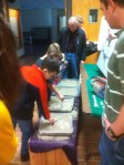
Walk Like a Dinosaur: We cut out and placed numerous anatomically-correct stenciled tracks of Tyrannosaurus, Dromaeosaurus, Titanosaur and Australopithicus (i.e., Lucy) to parallel a kraft-paper runway. On the runway, kids could strap little theropod feet onto their shoes, dunk them in some water, and then leave prints along the trackway. Kevin devised a fairly ingenious strap-on dino sandal made from velcro straps, plywood, and sponges for this. This was a huge hit and I was even able to wear the relatively tiny sponge feet, don Henry’s mache Velociraptor mask and scare the crap out of a 4yr old. Thanks to Dana Ehret for providing the inspiration for this and Ally for supervising to make sure everything went well.

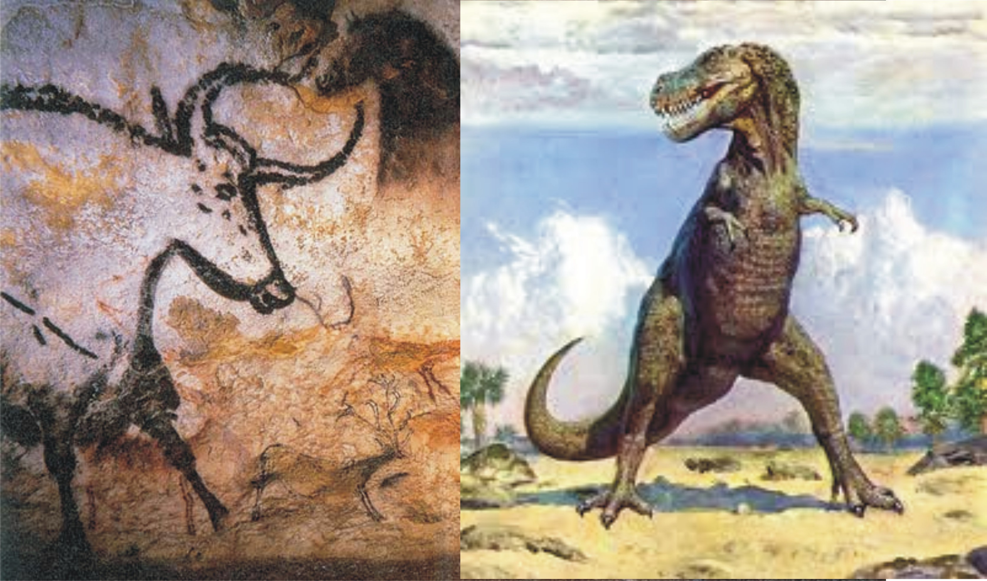
Little Lascaux: Kraft paper was hung on the wall. Stencils and examples of various Pleistocene animals that were depicted in the original cave were provided by Amy and Chet. Kids and parents and whomever was then enabled to create this massive mural on the wall. It included fire-breathing animals, rainbows, aurochs and potentially the earliest cave drawing of a hand turkey. We filled 2, 12ft murals with images that still sit rolled up in my office.

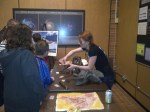
I Dig Dinosaurs: The venerable brush sand off a skeleton. Henry, Elizabeth and Ian sculpted a pretty sweet life-sized velociraptor skeleton out of ceramic clay placed it in 4 separate trays and covered it with vermiculite. We provided a poster of Velociraptor thanks to Scott Hartman as well as a copy of the Sue the T. rex quarry I still have from my younger days. This was a huge hit.

Know Your Knapping:  Alex Woods is a master knapper and held demonstrations all afternoon using a giant tool box full of stones, points, caribou antler, flint nodules, and atlatl. He always had a gaggle of kids around him as he pressure flaked, processed leather, and demonstrated how to take down a mammoth.

The Tables:

Everything Tyrannosaur: Casts, posters and help interpreting a random assortment of  tyrannosaur casts I have.

What is a Fossil?: trace fossils, molds and casts.

Cretaceous Fauna: We had some material from North America but mostly dinosaur casts I have from Gaston Design. So, this was a bit too weighted towards Asian dinosaurs. Next time we do this we’ll likely focus more on Missouri fauna and Geology, particularly if a recently submitted State grant funds us to revitalize the Missouri Chronister Dinosaur Site.

We Survived the Extinction: Basically a who’s who of vertebrate comparative material from the lab: alligators, turtles, birds, mammals, animals in jars, you name it. This was a very popular table led by Dave Dufeau and Kaleb Sellers.

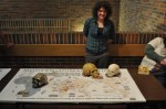
Neantertal’s vs Modern Humans: Anthro (Elizabeth, Rachel, Zack) had a nice couple of tables that had posters taped down onto the tables featuring biogeography, major anatomical features and an assortment of casts and artifacts illustrating the differences between the two arguable sympatric, hybridizing recent hominids.

Digital Paleo: Finally, we had a table that had a Clear & Stained alligator on a light box the got lots of attention. Next to it were two laptops. One had a loop of the Edmontosaurus chewing movie you can find at Palaeontologia Electronica and the 2nd had a draft our our 3D Alligator Jaw Muscle pdf that is in the pipeline at PLoS. It was great to see 8 year olds be able to easily manipulate the file to check out parts of the internal anatomy of an alligator.

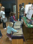
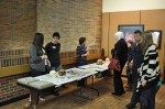
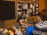
Below I will spam pictures from Scott Maddux and myself:


Posted in public education | 1 Comment

Dinosaurs & Cavemen Public Event

Saturday March 9th. Rock Bridge High School Planetarium. 1-5pm

Saturday March 9th. Rock Bridge High School Planetarium. 1-5pm

Saturday March 9th, a brigade of University of Missouri undergraduates, graduate students, and faculty will be at the local high school planetarium to provide an afternoon of discovery and excitement open to the general public. Come by, bring your families, catch a movie, help paint “Little Lascaux”, see how your footprints compare to those of human ancestors and Tyrannosaurus, help excavate fossils, and learn how global climate change has impacted plants and animals during the Late Cretaceous and Late Pleistocene time periods. Support your local planetarium! Support your local paleontologists!

March Public Shows at the Columbia Public Schools’ Planetarium
 
Earth’s Wild Ride” Event:
Dinosaurs, Cave Men, and Our Changing Planet
 
March 9, 2013
1:00pm, 2:00pm, 3:00pm, 4:00pm
 
Sixty five million years ago, an asteroid impact contributed to the extinction of the dinosaurs.  Twenty thousand years ago, much of a cooling Earth was covered in ice. These are just two ways Earth’s surface has changed over the millennia. 
 
Columbia Public Schools’ Planetarium & the University of Missouri’s Department of Pathology and Anatomical Sciences and Department of Physical Anthropology are teaming up to show just how our changing planet effected life on its surface.
 
·        Watch Earth’s Wild Ride planetarium show and see a tour of the night sky.
·        See dinosaur fossils
·        Compare your foot print to those of a dinosaur
·        Learn how cave men lived
·        Paint a cave mural
·        and more …
 
In the Planetarium:
Earth’s Wild Ride is set on the surface of the Moon in the year 2081.  A grandfather and his grandchildren watch a solar eclipse from scenic cliffs overlooking their moon colony. As they watch the Moon’s shadow move across Earth, the grandfather tells stories of crashing asteroids, roaring dinosaurs, frigid ice ages, erupting volcanoes, and more. A star tour will be give after each show.
 
Outside the Planetarium Doors:
Learn about dinosaurs and cavemen and the different times in which they lived. See fossils and artifacts! Help paint a cave mural! Compare your footprints with those of dinosaurs!
 
Admission to this event is free, but donations of $2 per person are welcomed.
 
CPS Planetarium is located within Rock Bridge High School at 4303 S Providence Rd in Columbia Missouri.  The planetarium is open for public shows on the second Saturday of each month.   School groups and private groups can reserve a weekday show by phone (573-214-3148) or email:Planetarium@columbia.k12.mo.us
Posted in public education | 1 Comment

A Brief History of Archosauriform Symphyses

Class-based evolution of the mandibular symphysis of archosauriforms. Holliday and Nesbitt, 2013.

Class-based evolution of the mandibular symphysis of archosauriforms. Holliday and Nesbitt, 2013.

Two years ago, I was invited to present in the Basal Archosaur symposium at the Latin American Congress of Vertebrate Paleontology in San Juan, Argentina. Holy cow do they know how to put on a good conference.  This symposium has turned into a Special Issue of the Geological Society of London. Papers in this volume will be trickling out over the next few months I presume. Ours is out now though. Sterling and I saw it as a good opportunity to begin a larger project on the functional significance and evolution of chins in archosaurs. Stemming from my earlier work on lizard symphyses (which we still have unpublished data of), ongoing work on crocodyliform chins, and a general interest in cranial arthrology, this paper presents the broad picture of diversity of symphyses among early archosaurs and up into the crown groups. From reading the paper, I hope people will take home just how incredibly diverse the chin, and feeding apparatus really is among this exciting group of vertebrates, and how often members of the larger group have experimented with convergent morphotypes. I think this is evidenced by the colorful character tree to the right (Fig. 12 from the paper).

One thing that I’ve been saying in the talks I give about this research that doesn’t appear strongly in the paper is that I’m not quite satisfied with the Scapino classification system as it applies to archosaurs and other reptiles. This system (Class I-IV) generally describes the overall morphology of the symphyseal plate–the ligamentous surface of the dentary that attaches to the other dentary. Using it was a good start since numerous mammal-centric papers use it to describe the joint. And I think it does an adequate job of broadly categorizing chins into unfused vs fused joints. But this does little to tell us about the internal architecture of fibers (which vary among animals, and may be woven, parallel, and/or distributed differently in the joint), the role Meckel’s cartilage has in the joint, good ole variation, and other features of the chin such as integument, dentition, the predentary, and even its complementing premaxilla. My brain shuts down a bit when I think about how to study not only the symphysis, which is complicated enough, but the symphysis+premax as a single functional unit. ow. So, we’re digging more deeply into the dark corners of characters to better describe all of this beautiful, functionally insightful anatomy (See Protosuchus figure) as well as more quantitatively explore the evolutionary and developmental patterns underpinning the joint over time.

Gorgeous MicroCT data of Protosuchus (MCZ 6727) and its sexy chin.

Gorgeous MicroCT data of Protosuchus (MCZ 6727) and its sexy chin.

Stay tuned for more symphyseal goodness in the near future. As for access to the paper, for now, email me if you want a copy.

Citation: Holliday CM, Nesbitt SJ. 2013. Morphology and diversity of the mandibular symphysis of archosauriforms. Geological Society, London, Special Publications v379. 17pp. doi 10.1144/SP379.2.

Abstract: Archosauromorphs radiated into numerous trophic niches during the Mesozoic, many of which were accommodated by particular suites of cranial adaptations and feeding behaviors. The mandibular symphysis, the joint linking the mandibles, is a poorly understood craniomandibular joint which may offer significant insight into skull function and feeding ecology. Using comparative data from extant amniotes, we investigated the skeletal anatomy and osteological correlates of relevant soft tissues in a survey of archosauromorph mandibular symphyses. Characters were identified and their evolution was mapped using a current phylogeny of archosauriforms with the addition of non-archosauriform archosauromorphs. Extinct taxa with the simple Class I condition (e.g., proterochampsids,rauisuchians”), rugose Class II (aetosaurs, protosuchians, silesaurids), and interdigitating Class III symphyses (e.g., phytosaurs, crocodyliforms) and finally fused Class IV (avians) build the joints in expected ways, though they differ in contributions of bony elements and Meckel’s cartilage. Optimization of the different classes of symphyses across a archosauromorph clades indicate that major iterative transitions from plesiomorphic Class I to derived, rigid Class II-IV symphyses occurred along the lines to phytosaurs, aetosaurs, a subset of poposauroids, crocodyliformes, pterosaurs, and birds. These transitions in symphyseal morphology also appear to track with changes in dentition and potentially diet.

Posted in Research News | 2 Comments

Transactions of the Royal Sounds of SVP

Yes, the seats squeaked upon sitting on them at this year’s Society of Vertebrate Paleontology meeting. During talks, most people sought to avoid the chirping by gently sliding laterally onto the cushion. But at the banquet, every applause was followed by waves of seat barks. Stay classy SVP!

All in all, the meeting was well executed, at a great location. Well done Host and Program Committees, and all the participants that made it possible.

Posted in academia, Research News, Uncategorized | Tagged | 3 Comments

Calls of the Wild

By: Ally McEntire

The calls of the baby gator in the Holliday lab got me thinking about alligator vocalizations. On a whim, I decided to look this up and found a little more than I had bargained for. Alligators and other crocodilians have a different vocal structure than any other reptile, amphibian or bird– all nearby relatives. Their vocal structure is actually quite similar to that of mammals. Instead of a syrinx like birds have, which involves air passing through the trachea while it vibrates at different rates; they have vocal folds—a larynx—just like humans.

This article details a study done on live juvenile crocs to study their vocalizations. It discusses a number of very specific things like sexual dimorphism in calls and amount of time the noises lasted. What I find more interesting than all of that is that their vocal structure is so unique. Even though they are evolutionarily related to these 3 other branches, their system of making noise is not really like any of their closest relatives. Basically, they have vocal folds, which contract and/or relax when the gator breathes out, like mammals.

This made me want to find out more about the ancestral line, if the present one is so estranged. Would other archosaurs resemble birds, lizards or crocs more closely? I’m not sure there’s a way to know this, considering the vocal cords are a soft tissue, and therefore incredibly difficult to preserve. But, if there were tissue attachments that could be identified, I think it’s something worth looking into.

Also, if you’re curious to hear our captive alligator making his own vocalizations, check Romer out here:

Romer Chirping

Posted in Uncategorized | Leave a comment

Reaching Out for Science

What is it that scientists do all day while they are watching their shrimp walk on a treadmill?  How do scientists know how Tyrannosaurus may have chewed or ran? How do they know if a molecule will work as a drug to target some disease? Scientists often find it difficult to translate their work’s importance to the public in a way they can understand without losing some of its content. Often “laypeople” are left confused with what looks like a waste of money in the name of big science.

So, how can the general public learn more about the inner workings of a research laboratory? Perhaps the key is to train student researchers early in their careers on how to write about science to the average person.

As a social experiment this year, members of the Holliday lab will try to bridge this gap by writing their own short pieces on relevant and interesting science.

By learning to translate often wonky, jargon-laden research to more palatable, engaging prose, lab members will hope to shed light on what happens in our lab, what questions we are asking, what discoveries we hope to make, and most importantly, why anyone should care.  

Of course, this should help the writers learn about science as well. Although the lab focuses on Vertebrate Functional Anatomy and Evolution, do not be surprised if other topics creep in. 

Posted in Uncategorized | Leave a comment

Ruminations on sauropsid cheeks

I noticed some FB flareup over dinosaur cheeks and had written all this in the box Facebook page and then decided it would be ludicrous to post so I moved it here…Apparently, dinosaur cheeks have become all the rage again in the blogosphere. Having only seen one published paper on the topic ever (Fabian Knoll’s 2008 paper) I’m still perturbed there aren’t other papers. But this region is way cool, apparently there is a new paper coming out on the topic. Being well aware of Witmer’s early work with Mike Papp on the cheeks vs freaks, Tobin H’s work on osteological correlates of skin (jeez I hope someone is pestering Tobin with a hot poker about this instead of just taking heavily from his published papers) and Ashley M’s still-evolving work on foramina…it is not an easy topic. My research on jaw muscles bumped into the buccinator/cheek/rictus problem alot, so I’m aware of the available evidence. (By the way, my favorite branch of the trigeminal nerve is the n. anguli oris—what’s your favorite CN V branch? Hmmm?)

 I hate the term cheeks as it always suggests the mammalian variety with a buccinator muscle (for which there is ZERO evidence for in saurpsids [yes, dinosaurs are also sauropsids!]).  The German anatomists (Luther, Lakjer, et al.) used either Mundplatte, and then later, the rictal plate, or rictus to describe the corner of the mouth in sauropsids (see my 2007 J Morph paper if you want more lit cited). Its rich with nerve endings, vasculature, glands etc and is easily seen in the corner of that snapping turtles mouth (see link above), crocodilian mouths, most birds. The rictus tends to directly cover the corner of the mouth where the temporal and  pterygoideus muscles cross paths as they attach to the mandible near the coronoid process, usually just underneath or caudal to the orbit.  Mammals have a rictus too– its the really sensitive edge of the cheek that lacks muscles but is packed with sensory receptors–go ahead, pinch it. Some snakes developed venom glands within the rictus (McDowell SB. 1986. The architecture of the corner of the mouth of colubroid snakes. J Herpetol 20:353–407.).

Anyway. I don’t see what’s so contentious aside from having a cutesy Dinocheek media event. Well endowed Mundplattes are not uncommon among sauropsids as people keep pointing out.  Why does there have to be one Dinosaurian bauplan for “cheeks” (ugh). Clearly with all the evolution that has happened in these groups, needing more/less skin over the mouth must have been necessary for chewing vs gape?  Clearly having a freaking giant osteoderm there lends cred to ankylosaurs having cheeks. but IMO, I never thought the old addage (err hypothesis?) “food would fall out of their mouth otherwise” to ever be a good approach to support the presence of skin there otherwise.  But perhaps there is upcoming evidence via SVP abstract to support it (yay!).  I feel like this whole debate is driven by paleoart, and not some true scientific endeavor (no offense artists-I understand the pains you often take to get things accurate). I sent most of this same information to Tylor Keillor, like 5 years ago to help with his sculptures-so its way cool to see his work progress that way it has been (see Project Dryptosaurus).

To me, a true cheek is when the rictus becomes decoupled from the true corner of the mouth (back near the coronoid process) and extends rostrally beyond to serve some function with the oral cavity.  So the cheekiest archosaur? My vote is the flamingo. Flamingos stretch the corner of the mouth past the corner of the mouth, past the orbit, past the antorbital cavity! past the transition from bony mandible to beaky mandible (holy crap!). And there is fairly clear function for this given the flamingo’s derived lingual pumping/filtration method of feeding. Here is an unpublished picture from our 2006 Anat Rec Flamingo Head Vessel paper.  Now that’s a @#$@# dinosaur cheek!

Flamingo extended rictus err dinosaur cheek

Posted in the best science is angry science, Uncategorized | 5 Comments

Travels to Rockefeller State Refuge

Alligator hanging out along Route 82

Rockefeller State Refuge is an expansive area of the western end of Louisiana’s swampy coast which prides itself as being one of the key DNR sites to aid in the rescue of American Alligators when they were endangered several decades ago (Link to Map and Location).  Today, besides maintaining a large wildlife management area full of birds, fish, herps, and sporting enthusiasts, they manage the region’s alligator population, work with commercial farmers and supply most of the alligators used in research in North America. If you didn’t already know, research in alligators is booming. There is strong interest in alligator and crocodilian genomics, hematology and disease resistance, biomechanics (for example…the death roll..), cardiopulmonary and developmental physiology, let alone our persistence in using them as a comparative anatomical model for vertebrate paleontology, functional anatomy and evolution.

Early morning Sun, Rockefeller Jan 31, 2011

 In February 2011, Henry Tsai and I drove down to collect alligator cadavers and made it back to Columbia at 3am, about 1hour before a blizzard hit and snowed us in for 2 days. It was a drive of legend in which we threaded the needle between two separate winter storms driving up through Western Arkansas. The alligators were quite comfy in the back of the truck, in my driveway while  we were snowed in.

The morning after picking up gators, after a 15hr drive through winter weather, Feb 1, 2011.

Needless to say, there are few contrasts in weather than experiencing sunrise over a balmy swamp one morning, and then 24 inches of snow the next.  The gators were used for research as well as a fairly popular high school workshop “Inside Alligators”we put on a week or so after we returned.

This June, I was accompanied by Ohio U/Witmer lab alum and current Mizzou Lecturer Dave Dufeau, and two undergraduates, Cortaiga Gant and Julie Tea. Cortaiga has been part of Project Gator Chin for a while whereas Julie is a Visiting Summer Fellow from University of Houston. This trip made for a good experience for them–none of whom had ever visited a place like Rockefeller and everyone got a chance to see, hold, and work with alligators. We stopped in Baton Rouge on the way down, stuffed our faces with shrimp and oysters at The Chimes and had a relatively leisurely drive down the coast the next day to get to Rockefeller. We then drove the entire way back to make sure the eggs and cadavers were taken care of.

Beach West of Cameron, LA

We visited Rockefeller this trip to load up a bunch of eggs we are currently incubating in the lab. Alligator eggs typically take about 65 days to hatch and are laid in a large, mother-guarded, mounded nest in late May/Early June. The Refuge staff use helicopters and airboats to identify and flag nests every season. They also collect recently-laid eggs and keep them in large outdoor incubators. The eggs are given to researchers or are hatched and raised until they are released into the wild or used otherwise.

Julie, Dave, and Cortaiga at Rockefeller, June 2012

Rockefeller has something different going on every time I visit. I’ve seen other students there collecting blood and gut bacteria. This time they had a number of alligator gar carcasses being prepped for someone’s study. Rockefeller received some serious damage from Hurricanes Rita and Katrina in 2005. One the “major” losses was their large outdoor, walk-in freezer, which had dozens of frozen specimens including the head of a gator that must have been pushing 10feet long…all swept away. None of this would be possible without the support and effort of Supervisor Ruth Elsey. Ruth has championed research and made the resources of the Refuge available to researchers and students from around the world. Ruth is always keen to help out people with research projects and educational materials and is always a welcoming host at Rockefeller.

Holding Gators

Alligator gar heads

Ruth showing Julie and Cortaiga a local gator nest. Momma is just under the reeds at the bottom of photo.

Posted in Research News | Tagged , , | Leave a comment

Cartilage Fusion in Gator Chins

Mandibular joints and Meckel’s cartilage in Alligator.

Summer in the Holliday Lab is getting exciting. A busy Spring has resulted in a couple new projects coming out later in the Fall (more later) but we’re deep in new directions in the lab including our first stint into Evo-Devo. Some students and I traveled to Louisiana to retrieve about 100 Alligator eggs to run an experiment this summer on the development of the Alligator chin. We’re keen to understand when and how the chin, or mandibular symphysis develops and pinpoint particular mechanisms and events that occur during its in ovo transformation.

Meckel’s cartilage is the cartilaginous rod that provides the scaffold for the ossifying mandible and 1st Branchial arch derivatives in the vertebrate skull. So, in reptiles, it promotes the ossification of the bones of the lower jaw and quadrate and remains as a persistent cartilage throughout life (Top Figure). This is in stark contrast to the mammalian condition in which the cartilage cavitates, aids in the formation of the dentary, 2 middle ear ossicles, and the tympanic ring. This caudal end of the system is better known-with questions involving the evolution and development of the mammalian ear driving many research directions. If you want to read more, I point you to the research of Abigail Tucker and  Zhe-Xi Luo, among many others.

Cross-section through suture and Meckel’s cartilage and CT-based 3D model of mandibular symphysis with bone, and without showing Meckel’s cartilage and sutural ligament.

At the rostral end of Meckel’s cartilage, out at the mandibular symphysis, we find animals doing a couple of different things. More often than not, the two cartilage rods (Left and Right) remain independent of one another and the symphysis is solely a membranous, ligamentous syndesmodial joint that fully fuses in primates, oviraptors, neoavians and a handful of other vertebrates, or may be sutured but unfused (dogs, crocodyliforms) (Middle Fig), or even mobile as in snakes, some lizards, possums and other animals probably. Second, research into the development of the human chin has found that the two cartilaginous rods stick together during development and eventually recede, occasionally leaving small nodular cartilages. Although the 2 cartilages stick to one another, the perichondral layer remains patent. We found a similar pattern in Iguana chins in our 2010 Anat Rec paper on lizard mandibular symphyses. Third, and now to the point. In alligators and geckos, Meckel’s cartilage not only sticks to its opposite member, but it obliterates the perichondral borders between the two cartilaginous rods, forming a continuous cartilaginous rod from the left articular all the way around the chin and back to the right articular.

That’s cool! Buy why? Is it adaptive? We’ve got some ideas. How? we’re working on it. Why do we care? working on that too 🙂 but in general, mandibular symphyses are important cranial joints functionally, so understanding their development and evolution are key goals if you want to understand the how the vertebrate head works. Meckel’s cartilage is often entwined in craniofacial defects that affect branchial arch development. Archosaur chins are really cool and quite diverse and thus may shed light on form, function, and ecology of feeding during their evolution.  Also, paleontologists have started to wiggle chin joints with more frequency these days, what with various animation and modeling applications,  despite there being what I consider a total lack of published research on how the joint is built (my fault I guess) and functions (in the works!)  in living reptiles.

Cleared and Double-stained alligator embryo showing the incipient double fusion and spatulate process of Meckel’s cartilage at the symphysis.

I digress… Fourth…Alligators take this cartilage fusion one step further in that a spatulate structure extends rostrally from the main body of Meckel’s cartilage  and forms a second fusion which eventually seals up with the caudal, main body of the cartilage. This spatulate form persists throughout life in alligators, leaving a large, flat trough within the bony symphysis. Is this fusion similar to those fusions we see in the hyoid skeleton, or the chondrocranium? probably. We’ll find out.

This summer we’re growing up Alligators to capture the time during development when this fusion occurs–I’ll post about that next.

Posted in Research News | Tagged , , , , | 4 Comments

SVP 2011 Report- Education & Outreach Poster

Yes, it’s been over a month since SVP met in Las Vegas. Shoot, I never blogged about my Argentina trip either. I’m so behind. I flew out during the day from Memphis, and being a clear day, I was able to basically lean my head on the window and watch Texas, New Mexico, and Arizona pass under us-always an amazing view. I took a few good pictures of the Grand Canyon from 30k ft up.

Grand Canyon from the plane

It was a good meeting, we had 2 Undergraduates and 2 PhD students give talks and a poster, and lab alumnus Nick Gardner also gave a great talk. I was really proud of all of their effort in making SVP go so well. To reward them, I punished them by taking them to the Banquet dinner and sitting through the awards ceremony. It was a horrifically sick joke, perhaps the meanest one I have ever subjected someone to. For that, I apologize.

SVP had some personally notable sessions including the Croc sessions (duh), the symposium on Laramidia, and the symposium on limb development. Of course there were interesting presentations scattered about the other sessions and posters.

Posters and orals presented by Holliday lab denizens were:

Gant CA, Skiljan BJ, Tsai HP, Folk B, Holliday CM. Alligators near and far: Using the Maps in Medicine: Inside Alligator high school workshop and 3D Alligator website as educational tools in anatomy and evolution.

Gardner NM, Bullar BA, Holliday CM, O’Keefe FR. Cranial anatomy in the basal diapsid Youngina capensis and its relevance to higher radiations of Permo-Triassic neodiapsida.

George ID, CM Holliday. Trigeminal nerve morphology in Alligator mississippiensis: Implications for inferring sensory potential in extinct crocodyliforms.

Holliday CM, Gardner NM. A new eusuchian crocodyliform with novel cranial integument and the origin of Crocodylia.

Skiljan BJ, Gant CA, Holliday CM. Structure and function of a protosuchian mandibular symphysis using anatomical insights from Alligator mississippiensis.

Tsai HP, Ward CV, Holliday CM. Pelvic anatomy of Alligator mississippiensis and its significance for interpreting limb function in fossil archosaurs

I’m happy to say that Ian’s paper on Alligator trigeminal nerve scaling is in review and the Shieldcroc paper is hopefully through its last minor revisions at PLoS ONE, so we should hopefully see those publications on the horizon. We’re currently developing Becci and Cortaiga’s work on Alligator and Protosuchian symphyses into papers.  The Shieldcroc presentation was selected by SVP to be a part of the media event. That made for some excitement before, during, and after the meeting. I’ll save my thoughts on that whole experience for when the actual paper comes out.

Among all these things, I had the most fun talking at the Outreach and Education posters which included a number of great methods, techniques, and ideas about how to broaden participation of various demographic groups in paleontology, anatomy and evolution education. We presented our own poster which served as a hybrid approach to how we are using Alligators as educational tools in regional High School education and Internet outreach. I’ve written about both of these events here previously: 3D Alligator; Inside Alligators. You can download a small version of the poster here.

Posted in Uncategorized | Tagged , , | Leave a comment