Class-based evolution of the mandibular symphysis of archosauriforms. Holliday and Nesbitt, 2013.
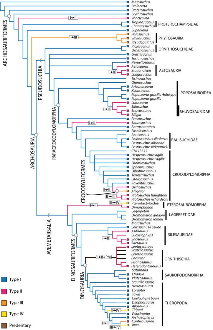
Two years ago, I was invited to present in the Basal Archosaur symposium at the Latin American Congress of Vertebrate Paleontology in San Juan, Argentina. Holy cow do they know how to put on a good conference. This symposium has turned into a Special Issue of the Geological Society of London. Papers in this volume will be trickling out over the next few months I presume. Ours is out now though. Sterling and I saw it as a good opportunity to begin a larger project on the functional significance and evolution of chins in archosaurs. Stemming from my earlier work on lizard symphyses (which we still have unpublished data of), ongoing work on crocodyliform chins, and a general interest in cranial arthrology, this paper presents the broad picture of diversity of symphyses among early archosaurs and up into the crown groups. From reading the paper, I hope people will take home just how incredibly diverse the chin, and feeding apparatus really is among this exciting group of vertebrates, and how often members of the larger group have experimented with convergent morphotypes. I think this is evidenced by the colorful character tree to the right (Fig. 12 from the paper).
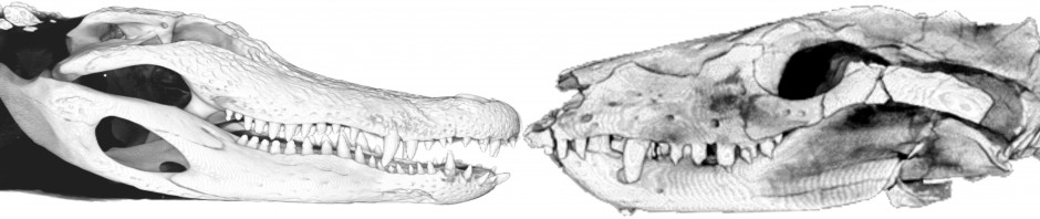
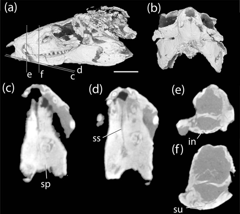
One thing that I’ve been saying in the talks I give about this research that doesn’t appear strongly in the paper is that I’m not quite satisfied with the Scapino classification system as it applies to archosaurs and other reptiles. This system (Class I-IV) generally describes the overall morphology of the symphyseal plate–the ligamentous surface of the dentary that attaches to the other dentary. Using it was a good start since numerous mammal-centric papers use it to describe the joint. And I think it does an adequate job of broadly categorizing chins into unfused vs fused joints. But this does little to tell us about the internal architecture of fibers (which vary among animals, and may be woven, parallel, and/or distributed differently in the joint), the role Meckel’s cartilage has in the joint, good ole variation, and other features of the chin such as integument, dentition, the predentary, and even its complementing premaxilla. My brain shuts down a bit when I think about how to study not only the symphysis, which is complicated enough, but the symphysis+premax as a single functional unit. ow. So, we’re digging more deeply into the dark corners of characters to better describe all of this beautiful, functionally insightful anatomy (See Protosuchus figure) as well as more quantitatively explore the evolutionary and developmental patterns underpinning the joint over time.
Gorgeous MicroCT data of Protosuchus (MCZ 6727) and its sexy chin.
Stay tuned for more symphyseal goodness in the near future. As for access to the paper, for now, email me if you want a copy.
Citation: Holliday CM, Nesbitt SJ. 2013. Morphology and diversity of the mandibular symphysis of archosauriforms. Geological Society, London, Special Publications v379. 17pp. doi 10.1144/SP379.2.
Abstract: Archosauromorphs radiated into numerous trophic niches during the Mesozoic, many of which were accommodated by particular suites of cranial adaptations and feeding behaviors. The mandibular symphysis, the joint linking the mandibles, is a poorly understood craniomandibular joint which may offer significant insight into skull function and feeding ecology. Using comparative data from extant amniotes, we investigated the skeletal anatomy and osteological correlates of relevant soft tissues in a survey of archosauromorph mandibular symphyses. Characters were identified and their evolution was mapped using a current phylogeny of archosauriforms with the addition of non-archosauriform archosauromorphs. Extinct taxa with the simple Class I condition (e.g., proterochampsids, “rauisuchians”), rugose Class II (aetosaurs, protosuchians, silesaurids), and interdigitating Class III symphyses (e.g., phytosaurs, crocodyliforms) and finally fused Class IV (avians) build the joints in expected ways, though they differ in contributions of bony elements and Meckel’s cartilage. Optimization of the different classes of symphyses across a archosauromorph clades indicate that major iterative transitions from plesiomorphic Class I to derived, rigid Class II-IV symphyses occurred along the lines to phytosaurs, aetosaurs, a subset of poposauroids, crocodyliformes, pterosaurs, and birds. These transitions in symphyseal morphology also appear to track with changes in dentition and potentially diet.

I love the caliber of this site, is truly fantastic to browse through
the content available right here.
How could I join your google+ page? Have one?